
Sani, Peyton and Dohnalkova reported in 2006 that the toxic effects of uranium(VI) were studied using Desulfovibrio desulfuricans G20 in a medium containing bicarbonate or 1, 4-piperazinediethane sulfonic acid disodium salt monohydrate (PIPES) buffer (each at 30 mM, pH 7). Uranium(VI) toxicity was dependent on the medium buffer and was observed in terms of longer lag times and in some cases, no measurable growth. The minimum inhibiting concentration (MIC) was 140 µM U(VI) in PIPES buffered medium.
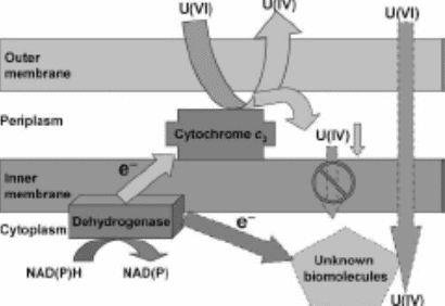
The above figure illustrates the Uranium detoxification model in Desulfovibrio desulfuricans G20. NAD(P)H reduced form of nicotinamide adenine dinucleotide phosphate; U(IV) reduced uranium; U(VI) uranium(VI). This is 36 times lower than previously reported for D. desulfuricans. In all cases in which G20 grew in the presence of U(VI), the final cell protein yield was equivalent to that of the U(VI)-free control. In 24 h, D. desulfuricans G20 (40 mg/L total cell protein) removed 50 µM U(VI) from solution in PIPES buffer as compared to 96 µM U(VI) in bicarbonate buffer under anaerobic, nongrowth conditions. Even though the solubility of U(VI) was significantly lower in PIPES buffer than in bicarbonate buffer, U(VI) was much more toxic in PIPES buffer than in bicarbonate buffer.
Analysis of thin sections of G20 treated with 90 µM U(VI) in medium containing PIPES buffer revealed that only a very small fraction of cells had reduced uranium (U) precipitates in the periplasmic spaces. In the presence of bicarbonate buffer, however, reduced U was observed not only in the periplasm, but also in the cytoplasm.
Selected area electron diffraction patterns and crystallographic analysis of transmission electron microscope lattice fringe images confirmed the structure of precipitated U in the cell periplasm and cytoplasm as being that of uraninite. These results suggest that U(VI) toxicity and the detoxification mechanisms of G20 depend greatly on the chemical forms of U(VI) present.
Managed & Built by I2M Consulting, LLC | © 2010-